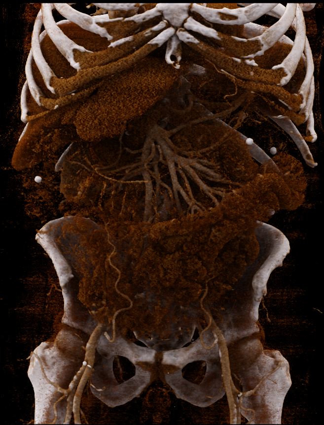
Lior Molvin joins Bill and Kristan for a discussion of both CT and MRI studies of the small bowel.
Show Notes
- Describe the process of a radiographic study of the small bowel.
- Name specific improvements to CT technology which contributed to enabling CT exams of the small bowel.
- List the stages of contrast enhancement in a CT exam of the small bowel.
- Describe the characteristics of the various types of oral contrast agents for both CT and MR Enterography studies.
- Describe the patient prep prior to a CT Enterography exam.
The content of this Contrast Enhanced MRI Program is intended for healthcare professionals who work in the MRI environment. These individuals include MRI Technologists, Imaging Nurses, MRI Researchers, and Others.
This program has been submitted for 1.00 hours of Category A CE credit as designated by the International Society for MR Radiographers & Technologists (ISMRT) RCEEM.
Release Date: 11/08/2024 | Expires: 11/08/2026
Participants may claim only one type of credit for this educational activity.
Faculty and Planner Disclosures:
As an accredited provider of continuing medical education, Northwest Imaging Forums, Inc. (NWIF) ensures balance, independence, objectivity, and scientific rigor in all its educational activities. In accordance with the ACCME Standards for Integrity and Independence in Accredited Continuing Education, all faculty and planners are required to disclose all financial relationships with ineligible companies within the last 24 months. All relevant financial relationships have been mitigated by NWIF to ensure the integrity of the CME activity. The planner has nothing to disclose.
Course Content
Podcast Content
Episode 37 Enterography – Evaluation
You don't currently have access to this content
Episode 37 Enterography – Quiz
You don't currently have access to this content